INTRODUCTION
Ampullary carcinoma is defined as an epithelial malignant neoplasm arising from the epithelium lining the ampulla of vater located at the junction where the common bile duct (carrying bile from the liver and secretions from the pancreas) empties into the duodenum (upper small intestine) or the immediate surrounding duodenal mucosa This cancer can cause obstruction of the common bile duct and prevent bile from flowing into the intestine and out of the body. Adenocarcinoma of the ampulla of Vater is a relatively uncommon tumor. A case of ‘Ampulary Carcinoma is hereby presented.
CASE HISTORY
A 60 years old lady presented with yellowish discoloration of eyes and urine for three months associated with itching. There was significant weight loss of about 25% in last 3 months. She was a chronic smoker for last 40 years. There is no history of alcoholism or any other significant past or family history.
CASE FINDINGS
General condition was fair with icterus, palpable inguinal and axillary lymph nodes, and no pallor. Abdomen was soft and non-tender. No organomegaly was seen. Investigations showed liver function tests as Total bilirubin- 11.0mg/dl, Direct (conjugated) bilirubin- 09.7mg/dl, Indirect (Unconjugated) bilirubin 01.3mg/dl, ALT- 70 IU/L (< 40 IU/L), AST- 74 IU/L (< 40 IU/L) and Alkaline Phosphatase- 1109 (110-310 IU/L), Hemoglobin- 10.9 gm/dl and ESR- 105 mm/hr. Preoperative USG findings showed dilated CBD and gall bladder, and mass protruding into the duodenal lumen with intact mucosa. Patient underwent Whipple’s procedure.
PATHOLOGICAL FINDINGS
Gross
-
Specimen consists of distal part of the stomach, duodenum, part of the pancreas and terminal portion of bile duct.
-
Cut section of the duodenum shows a small firm tumor measuring 1.5 x 1.3 x 1 cm.
-
The tumor has tiny papillary projections below the duodenal mucosa near the opening of the ampulla of Vater. The duodenal mucosa over the growth appears uninvolved by the tumor.
-
Gall bladder and stomach look unremarkable.
Microscopic findings
-
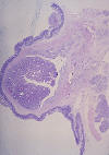
Section from the tumor shows tissue lined with normal duodenal mucosa. A tumor is seen arising from the base of the polypoid nodule below the submucosa (Figure 1). The tumor has the appearance of villoglandular polyp formed by crowded glands lined by benign cells and with superficial papillary areas (Figure 2). At places there are malignant changes showing glands lined by malignant cells with pleomorphic, vesicular nuclei and marginated chromatin and moderate amount of cytoplasm. There are areas with extracellular mucin collection. The submucosa of the duodenum shows infiltration by the same malignant cells and collection of extracellular mucin (Figure 3).
-
Section from the stomach and pancreas is normal and unremarkable.
-
Proximal and distal surgical margins are free of tumor.
-
Lymph node shows reactive changes.
Figure 1 |
Figure 2 |
Figure 3 |
HISTOPATHOLOGICAL DIAGNOSIS
Ampullary carcinoma- Grade II (80 % glandular patterns)- Size 1.5 x 1.3 x 1 cm- Intra ampullary carcinoma- Papillary type. It is invading the duodenal wall with all surgical margins free of tumor. No lymph node involvement is seen. There is no evidence of lymphatic, perineural or vascular invasion.
Tumor is arising in villous adenoma, pTNM: pT2 pN0 pMX, Stage - II. It accounts for 0.2% of gastrointestinal malignancies. Depending upon the location it is classified as intra-ampullary, periampullay and mixed type. Microscopically this tumour is adenocarcinoma. Few cases arise from benign villous adenoma or villoglandular polyp. Metastases to regional lymph nodes, liver, lung peritoneum are noted. Poor prognostic factors are high stage, tumour size >2.5 cm, perineural invasion, angiolymphatic invasion, invasion of muscle of sphincter of Oddi, nodal metastasis, signet ring histology, and poor differentiation.


