Received: March 09, 2011
Accepted: May 01, 2011
Ref: Kachewar S, Bhadane S, Kulkarni D, Sankaye S. Giant peripheral osteoma of the mandible. Internet J Med Update. 2012 Jan;7(1):66-9.
GIANT PERIPHERAL OSTEOMA OF THE MANDIBLE
Sushil Kachewar* MD, DNB, Sushant Bhadane** MD, Devidas Kulkarni† MD and Smita Sankaye‡ MBBS
*Associate Professor, †Professor, Department of Radiology, ‡Resident, Department of Pathology, Rural Medical College, Loni, Maharashtra, India
**Consultant Radiologist, Nashik, Maharashtra, India
(Corresponding Author: Dr. Sushil Kachewar, Associate Professor, Department of Radiology, Rural Medical College, Pravara Medical Trust, PIMS (DU), Loni 413736, Maharashtra, India; Mobile: 0091-9921160357; Email: sushilkachewar@hotmail.com)
ABSTRACT
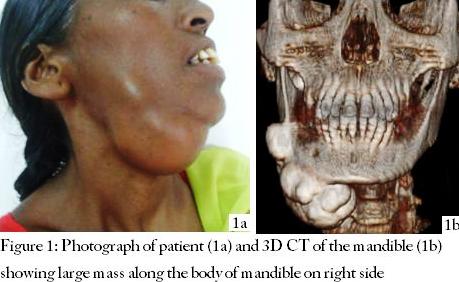
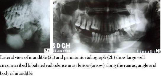
Osseous expansion of any body part is an unwelcome guest and deep are its impacts when it is located on the face. The bigger the lesion, the more bitter is the psycho-social trauma to the affected individual. This article describes the case of a 50 year old female who presented with painless swelling of the right submandibular region manifesting as a dreadful cosmetic disfigurement. The mass had been progressing slowly for the last 15 years. Imaging showed a giant peripheral osteoma of 10.8 cm involving buccal and lingual surface of the body, ramus, angle and inferior border of the right side of mandible. To the best of our knowledge, a giant peripheral osteoma of mandible having size more than 10 cm has never been reported earlier.
KEY WORDS: Giant peripheral osteoma; Swelling of mandible; CT scan, Panoramic view
I
| Figures |
|---|
 |
 |
 |
 |