INTRODUCTION
Tuberculosis continues to be a major health hazard, inspite of notable advances in its diagnosis and treatment [1]. This systemic disease presents with varied clinical manifestations as pulmonary tuberculosis or extra pulmonary tuberculosis.
Extra pulmonary tuberculosis accounts for almost 15% of all cases of tuberculosis. Among extra pulmonary form, splenic tuberculosis is exceptionally rare clinical condition. This form of tuberculosis is normally seen as a part of miliary tuberculosis and is rarely the isolated entity or presenting feature. Here we are reportingting a case of splenic tuberculosis which presented as an isolated entity.
CASE REPORT
A non diabetic, non hypertensive immunocompetent middle aged woman from good socioeconomic background presented with intermittent low grade fever, pain in left hypochondriac region and progressive weight loss for about 2 months. On examination she was pale and afebrile. Per abdomen examination revealed moderately enlarged and tender palpable spleen. Routine blood investigations i.e. haemogram and chest X ray were normal except that ESR was found to be raised (50mm/hr Wintrobe’s method). Blood culture was negative. Ultra sound of abdomen revealed multiple hypo-echoic lesions in an enlarged spleen, while CT scan showed diffuse lesions in the spleen. On the basis of radiological investigations provisional diagnosis was made as mass in spleen, possibly malignant. Splenectomy was carried out.
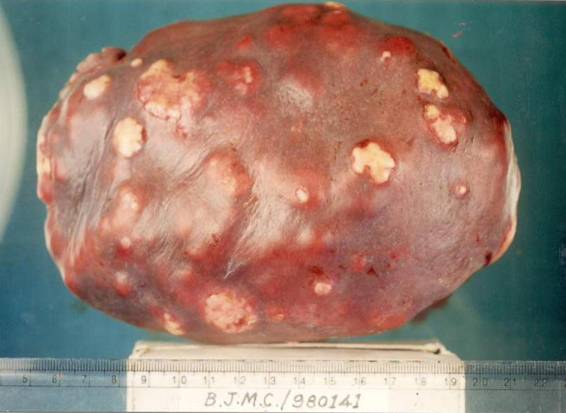
On gross examination of specimen multiple whitish nodules rising above the surface of about 1-3 cm in size were noted on the external surface (Figure 1). Cut section showed multiple nodules coalescing to form large yellowish white mass of firm consistency (Figure 2). Hemotoxilin and eosin stained section of nodule showed large areas of caseation surrounded by multiple granulomas of epitheloid cells and Langhan’s giant cells throughout the splenic pulp. Surrounding splenic parenchyma was within normal limits. However acid fast staining of section did not show presence of acid-fast bacilli.
 |
 |
Figure 1: Multiple whitish nodules on the External Surface |
Figure 2: Cut section showing large yellowish white mass |
A biopsy from the diseased portion of spleen was sent to Microbiology department for further follow up. The Specimen was processed for microscopy, culture and polymerase chain reaction to rule out possibility of Mycobacterial infection. The received tissue was minced in sterile saline. Minced tissue was used to prepare smears which were stained by standard Zeil Nelson staining technique. Small portion was inoculated on Lowenstein Jensen medium and incubated at 370C. The remaining tissue was subjected to standard phenol chloroform DNA procedure [2]. The extracted DNA was subjected to amplification. PCR was carried out using IS 6110 insertion sequence based primers giving 123bp product. An initial denaturation was done at 950C for 3min. to ensure complete separation of two templates. Amplification was carried out as follows; denaturation at 940C for 40sec, annealing at 650C for 40sec and extension at 720C for 40sec. A final extension cycle of 720c for 4min was performed to ensure complete extension of partially extended PCR product. A total of 30 cycles of amplification were done [3]. On amplification, product was visualised by gel electrophoresis. The amplified product showed 123bp product indicative of presence of mycobacterium tuberculosis infection (Figure 3).
 |
At the end of 5th week of incubation, Lowenstein Jensen medium showed growth of acid-fast bacilli. The isolate was subjected to a battery of biochemical tests. On the basis of results of biochemical tests, the isolate was identified as M. tuberculosis. Patient was put on antituberculous drugs. With splenectomy and antitubercular treatment patient showed improvement at the end of the 5th week in terms of weight gain and decreased ESR.
DISCUSSION
Clinically Tuberculosis may present as pulmonary or extrapulmonary disease. Of all the organs, lungs are the predominantly affected organs. Involvement of spleen in tuberculosis occurs in miliary/disseminated form of the disease. However isolated splenic tuberculosis or solitary tuberculosis of spleen is very rare in abdominal organs. When spleen is involved as an isolated organ, patient may have solitary tuberculosis or tubercular abscess. Splenic abscess is a comparatively commoner stage than the solitary or nodular stage when patient seeks medical advice.
Many reported cases of splenic tubercular abscess are found to have underlying HIV infection also [1, 4]. Splenic involvement was thought to be seen only in immunocompromised stage. However, there are sporadic case reports of splenic tuberculosis, mainly the splenic abscess where patient is immunocompetent [5]. Adil A et al [6] reported a series of 10 immunocompetent individuals with splenic tuberculosis. All of them had at least one site or organ affected by tuberculous infection. The common presenting clinical features are pyrexia of unknown origin and thrombocytopenia. Rarely, it has also been diagnosed incidentally during laparotomy that was carried out for abdominal trauma [7].
In the present study patient was immunocompetent. She neither had history of tuberculosis nor evidence of tuberculosis in any other organ. There was also no history of trauma. She came with the complaints of pain in hypochondriac region, low grade fever and weight loss. Diagnosis has been made accidentally during laparatomy which was carried out for suspected malignancy.
A case of splenic tuberculosis presented with weight loss and fever but no hypochondriac pain was reported by Ho PL et al [8]. A case of several finger tip sized palpable bilateral inguinal lymphnodes along with high grade fever and weight loss was finally diagnosed to have splenic tuberculosis. Although histopathologic examination of lymphnode was nonspecific for tuberculosis, acid fast bacilli could be demonstrated in the biopsy of splenic nodule in this case [9].
Diagnosis of isolated splenic tuberculosis is difficult and often delayed because of vague clinical manifestations. In almost all the reported cases diagnosis was made by radiologic examination followed by pathologic examination of fine needle aspiration, splenic biopsy or of splenectomy specimen. In our case ultrasound examination revealed hypoechoic lesions while CT scan demonstrated hypodense area in the spleen. However similar radiological picture is also seen in patients having fungal infection or malignancy. Radiology cannot pinpoint the underlying etiology. Therefore histopathological examination is necessary for etiological diagnosis. Histopathologically tuberculous infection can be identified by typical caseation along with granuloma of epitheloid cells and Langhans giant cells but it cannot differentiate whether infection is due to mycobacterium tuberculosis or atypical mycobacteria. If it is due to atypical mycobacteria, patient may not respond to the routine antituberculous drugs. In addition to this histopathological report is usually available by the third day from the date of submission of tissue. In the present study therefore, attempt has been made to confirm radiological diagnosis by microbiological investigation with the advent of technology, and identification of etiological agent up to the species level was done by Polymerase Chain Reaction.
Carry Mullis in 1985 for the first time demonstrated in-vitro amplification of DNA [10]. The technique slowly developed and shifted from research laboratory to diagnostic clinical microbiology laboratory. The main fascinating advantage of PCR is its rapidity and sensitivity with which results are made available. In the present case also the underlying cause of vague clinical symptoms could be identified within few hours of submission of specimen after splenectomy.
CONCLUSION
There are hardly any case reports of isolated solitary splenic tuberculosis where microbiological examination is carried out. This is one of the rare cases of isolated splenic tuberculosis in an immunocompetent individual where rapid diagnosis was achieved by PCR and confirmed by histopathology as well as culture. Although splenic tuberculosis is rare, it should be included in the differential diagnosis of PUO with splenomegaly regardless of the HIV status of the patient.
REFERENCES
- Prathmesh CS, Tamhankar AP, Rege SA, et al. SplenicTuberculosis and HIV-1 infection. Lancet. 2002;369(26):353.
- Sambrook J, Frisch EF, Maniatis T. Molecular Cloning: A laboratory manual vol. II, 2nd edition. Cold Spring Laboratory press. 1989.
- Eisenach KD, Crawford JT, Bates JH. Repetitive DNA Sequences as probes for Mycobacterium tuberculosis. Journal of clinical Microbiology. 1988;26(11):2240-5.
- Thomson SR, Ghimenton F. Splenic Tuberculosis. Postgrad Med J. 1999;75(888):578.
- Chandra S, Srivastava DN, Gandhi D. Splenic tuberculosis : an unusual sonographic presentation. Int J Clin PRact. 1999 Jun;53(4):318-9.
- Adil A, Chikhaoui N, Ousehal A, et al. Splenic Tuberculosis Apropos of 12 cases. Ann Radiol (Paris). 1995;38(7):403-7.
- Singh B, Ramdial PK, Royeppen E, et al. Isolated splenic tuberculosis. Trop Doct. 2005 Jan;35(1):48-9.
- Ho PL, Chim CS, Yuen KY. Isolated Splenic Tuberculosis presenting with pyrexia of unknown origin. Scand J Infect Dis. 2000;32(6):700-1.
- Sato T, Mori M, Inamatsu T, et al. Isolated splenic tuberculosis. Nippon Ronen Igakkai Zasshi. 1992 Apr;29(4):305-11.
- Mullis KB. The unusual origin of the Polymerase Chain Reaction. Scientific Americans. 1970;262(Apr):56-65.